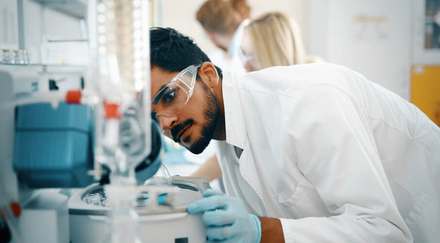
 Microfluidics chambers have a variety of uses. This article will cover the basics of using a microfluidics chamber. The sample preparation process involves the introduction of a sample through the microfluidics chamber. The fluids used in the chamber include HBSS, 1% human serum albumin, 1 mM CaCl2, and E-selectin. The sample preparation procedure also requires the use of a microfluidics device that can measure cellular behavior in real time. Flow through microfluidics chambers is governed by the Reynolds-averaged Navier-Stokes partial differential equations. The numerical solution of these equations can be achieved using a finite element method. This computational technique was used to analyse the flow within the microfluidics circuit. In order to simulate this method, the computational domain was discretized. This method allows for easy comparison of different flow parameters. Click for more details on microfluidics chambers on this website. A custom microscope was used to monitor the effect of photoperiod on protoplast growth. Microfluidic samples were inserted into the microscope and continuously observed for 80 hours. During the absence of light, the process of tip elongation was strongly inhibited. Growth restarted after a short lag. Continuous illumination resulted in globally constant growth. There are several advantages of microfluidics as a tool for studying plant development. A microfluidics chamber is a device that enables researchers to study and control liquid flow. Its construction involves a plastic container with multiple chambers. Its dimensions make it possible to easily manipulate and adjust the flow rate within the chamber. In this way, scientists can design and develop custom microfluidic devices. These devices will improve the way scientists test and design microfluidic systems. There are numerous benefits to using microfluidics chambers, and you can even design your own. The microfluidic chambers are cleaned with a hydrophilic block-polymeric surfactant solution prior to experiments. This solution contains 0.2 wt% of Pluronic F68 in water. Then, you can use them for long-term culture conditions. The chambers are compatible with the biomedical applications that your research requires. The microfluidic chambers chamber is able to facilitate a variety of processes, from cell culture to imaging. The microfluidic chip is made up of a single layer of flow-through microfluidic traps. Protoplasts can be immobilized in the microfluidic traps for further study. The microfluidic chip is portable and can be inserted into an incubator that controls illumination and record the development process continuously. You can then remove the microfluidic chip and perform the experiment again with a fresh medium. For more details related to microfluidic chambers, click here now! Microfluidics chambers are typically connected to side microchannels. These side channels serve many purposes, including delivering nutrients, bacteria, or viruses to cells. Besides serving these functions, microfluidics chambers can also be used to manipulate the cells mechanically and study their interactions. Some microfluidics chambers are so small that they can be connected together into an organ-on-a-chip system. One example of a biofluidics chamber involves measuring the surface densities of recombinant E-selectin. To determine these surface densities, an antibody conjugated to the E-selectin surface was coated onto the chamber. It was then incubated with antibodies for an hour. Free antibodies were removed by removing them with HBSS and fluorescence images were captured using a wide-field fluorescence microscope setup. Using a microfluidics chamber, the surface densities of the E-selectin were calculated by comparing fluorescence intensities of the E-selectin-coated microfluidic surface with those of single antibodies. Check out this blog to get enlightened on this topic: https://en.wikipedia.org/wiki/Organ-on-a-chip.
0 Comments
Leave a Reply. |
 RSS Feed
