|
5/13/2022 0 Comments Microfluidics Chamber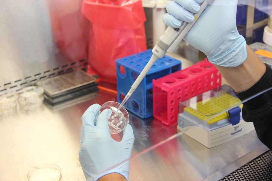 A microfluidics chamber is a device that combines fluid and mechanical properties. It is often used for cell culture experiments. This device is made of a chip with holes for the injection and removal of liquids. Active systems that pump liquid into and out of the microfluidic chamber include a pressure controller and a syringe-pump. Passive methods include hydrostatic pressure. These techniques allow scientists to test a wide range of molecules in a single chip. A microfluidics chamber has two main approaches to sample introduction. The first involves pumping a sample into the main channel and switching the flow to inject the sample into the second channel. Both methods rely on continuous deflection of particles in laminar streams. A physical barrier is often used to prevent particles from entering or exiting the chamber. Both methods have their advantages and disadvantages. Click here for more details on how you can contact your local laboratory expert. Another method involves the use of traps. Usually, traps are U-shaped with two lateral openings. This allows the culture medium to flow easily through them without generating dead volumes. It is also easy to tune the traps to achieve optimal growth. Various factors can help improve the efficiency of the traps. The trapping efficiency can be optimized by varying the cell density and the time of loading. Moreover, it has an advantage over other microfluidics systems. A microfluidic device is made with a filtration network that restricts fluid flow to specific regions of the substrate. A microfluidic master device can be created in as little as 15 minutes, depending on the size of the sample. The fabrication time of a twelve mm-long wall takes about 25 minutes. Creating a six-mm-long wall can be done in about 20 minutes. Alternatively, the two-photon fluorescence image of a microfluidic chamber is shown in Figures 1 and 2. The microfluidic chamber was set up with a sticky-Slide VI 0.4. Then, the chamber's inlet and outlet were connected to male Luer connectors. The cells were incubated in a sticky-Slide VI 0.4. In addition, the rolling buffer contained 1% human serum albumin. In addition, a programmable syringe pump was used for the experiments. You can learn more now here to understand the basics of microfluidic master device. The researchers then used an inverted optical microscope to capture the cell rolling behavior. They used a 20x objective to capture the image of the cell-rolling behavior. The microfluidic chamber was mounted on an inverted microscope, which was equipped with a 20x objective. A CCD camera was used to record the images. Lastly, the researchers incorporated a micropost array into the gradient generator to detect unwanted thrombi formation. The platform was also compatible with selective plane illumination microscopy (SPM) to study the interaction between HSPCs and the endothelial cells. Using a microfluidics chamber, scientists can easily measure the surface densities of recombinant E-selectin. This technique can be used for cell-rolling assays to identify which molecules play the role of the immune system in inflammatory response. If you want to know more about this topic, then click here: https://en.wikipedia.org/wiki/Microfluidic_cell_culture.
0 Comments
Leave a Reply. |
 RSS Feed
